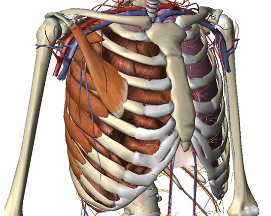
Anatomy Of Chest And Ribs : Rib Cartilage Injury Masnad Health Clinic / The sternum is also known as the breastbone.. The anatomy of the human ribs is made up of 24 ribs which are parted in 12 pairs (each on the left and right side of the chest wall), with the sternum, metasternum(the xiphoid process), and the costal cartilages all situated at the anterior of the chest wall, followed by the thoracic vertebrae on the posterior of the chest wall. Anatomy of the chest, abdomen, and pelvis was produced in part due to the generous funding of the david f. As part of the bony thorax, the ribs protect the internal thoracic organs. In humans and other hominids, the thorax is the chest region of the body between the neck and the abdomen, along with its internal organs and other contents. The cartilage strips are called costal cartilage (costal is the anatomical adjective that refers to the rib) and connect on their other end to the sternum.
In some patients an extra joint is seen in the anterior part of the first rib at the point where the bone meets the calcified cartilageneous part (arrow). Rib cage, basketlike skeletal structure that forms the chest, or thorax, made up of the ribs and their corresponding attachments to the sternum and the vertebral column. Anterior chest wall showing muscular attachments and neurovascular structures ribs 3 through 9 are typical ribs as described earlier while ribs 1, 2, 10, 11, and 12 are atypical. The word sternum originates from the ancient greek word 'sternon', meaning chest. The first seven ribs progressively increase in length, the lower five ribs then begin to decrease in length.
Slipping Rib Syndrome And Other Causes Of Chest Wall Pain Springerlink from media.springernature.com There are twelve pairs of ribs, all of which articulate with the vertebral column. However, only seven have a direct articulation with the sternum. Powerful muscles that move the head and arms attach to these bones as well. This article focuses on the unique structural characteristics in the internal thoracic diameters. The ribcage contains 12 ribs total on each side, divided into three different types. Anatomy of chest and ribs / the thoracic cage anatomy and physiology i. In humans and other hominids, the thorax is the chest region of the body between the neck and the abdomen, along with its internal organs and other contents. At the chest, many rib bones connect to the sternum via costal cartilage,.
Learn about chest anatomy with free interactive flashcards.
The rib below that is rib 2, and it connects to the t2 thoracic vertebra, and so on. The ribcage contains 12 ribs total on each side, divided into three different types. Ribs are highly vascular and trabecular with a thin outer layer of compact bone. We think this is the most useful anatomy picture that you need. Ten of the twelve ribs connect to strips of hyaline cartilage on the anterior side of the body. Anatomynote.com found chest bone, ribs, lung, heart, xiphoid process, sternum anatomy from plenty of anatomical pictures on the internet. As with all parts of the body, the anatomy and physiology of the chest wall are intimately intertwined. In some patients an extra joint is seen in the anterior part of the first rib at the point where the bone meets the calcified cartilageneous part (arrow). For more anatomy content please follow us and visit our website: They are extremely light, but highly resilient; As part of the bony thorax, the ribs protect the internal thoracic organs. Anatomy of chest and ribs. It is a flat bone that articulates with the clavicle and the costal cartilages of the upper 7 ribs (true ribs), while the 8th, 9th and 10th ribs (false ribs) are indirectly attached with sternum via costal cartilage of the ribs above.
Each pair is numbered based on their attachment to the sternum, a bony process at the front of the rib cage which serves as an anchor point. 2 joints between heads of the ribs and bodies of vertebrae (corresponding and upper). Ribs are highly vascular and trabecular with a thin outer layer of compact bone. The rib cage is a bony structure found in the chest (thoracic cavity). However, only seven have a direct articulation with the sternum.
Costochondritis Chest Wall Pain Rib Injury Clinic from www.ribinjuryclinic.com Each pair is numbered based on their attachment to the sternum, a bony process at the front of the rib cage which serves as an anchor point. The rib cage also anchors the bones of the head, neck, shoulders, and arms to the trunk of the body. Both the liver and the stomach are located in the lower chest region under the thoracic diaphragm, a sheet of muscle at the bottom of the rib cage that separates the chest cavity from the abdominal. Powerful muscles that move the head and arms attach to these bones as well. 2 joints between heads of the ribs and bodies of vertebrae (corresponding and upper). Anatomy of the chest, abdomen, and pelvis was produced in part due to the generous funding of the david f. Ribs eight to ten are the false ribs and are connected to the sternum indirectly via the cartilage of the rib clinical notes. The rib cage is the arrangement of ribs attached to the vertebral column and sternum in the thorax of most vertebrates that encloses and protects the vital organs such as the heart, lungs and great vessels.
It is made up of 12 pairs of ribs.
For more anatomy content please follow us and visit our website: The anatomy of the human ribs is made up of 24 ribs which are parted in 12 pairs (each on the left and right side of the chest wall), with the sternum, metasternum(the xiphoid process), and the costal cartilages all situated at the anterior of the chest wall, followed by the thoracic vertebrae on the posterior of the chest wall. 2 joints between heads of the ribs and bodies of vertebrae (corresponding and upper). Rib cage, in vertebrate anatomy, basketlike skeletal structure that forms the chest, or thorax, and is made up of the ribs and their corresponding attachments to the sternum (breastbone) and the vertebral column. 4, brachiocephalic vein (left side). Anterior chest wall showing muscular attachments and neurovascular structures ribs 3 through 9 are typical ribs as described earlier while ribs 1, 2, 10, 11, and 12 are atypical. The cartilage strips are called costal cartilage (costal is the anatomical adjective that refers to the rib) and connect on their other end to the sternum. The first rib is a short, flat rib that is much wider and more curved than those previously described. There are twelve pairs of ribs, all of which articulate with the vertebral column. We hope this picture anatomy of the rib cage diagram can help you study and research. The sternum is also known as the breastbone. The chest anatomy includes the pectoralis major, pectoralis minor and the serratus anterior. The rib cage is a bony structure found in the chest (thoracic cavity).
It is a flat bone that articulates with the clavicle and the costal cartilages of the upper 7 ribs (true ribs), while the 8th, 9th and 10th ribs (false ribs) are indirectly attached with sternum via costal cartilage of the ribs above. Anatomy of chest and ribs : The ribs are a set of twelve paired bones which form the protective 'cage' of the thorax. The bones of the chest and upper back combine to form the strong, protective rib cage around the vital thoracic organs such as the heart and lungs. However, only seven have a direct articulation with the sternum.
Chest Wall Radiology Key from i0.wp.com For more anatomy content please follow us and visit our website: For more anatomy content please follow us and visit our website: Anatomynote.com found chest bone, ribs, lung, heart, xiphoid process, sternum anatomy from plenty of anatomical pictures on the internet. The cartilage strips are called costal cartilage (costal is the anatomical adjective that refers to the rib) and connect on their other end to the sternum. As with all parts of the body, the anatomy and physiology of the chest wall are intimately intertwined. Each pair is numbered based on their attachment to the sternum, a bony process at the front of the rib cage which serves as an anchor point. Anatomy of chest and ribs : To carry out the unique functions performed by the chest wall, the anatomic structures are formed precisely for maximal efficiency.
As part of the bony thorax, the ribs protect the internal thoracic organs.
As part of the bony thorax, the ribs protect the internal thoracic organs. At the chest, many rib bones connect to the sternum via costal cartilage,. Anatomynote.com found anatomy of the rib cage diagram from plenty of anatomical pictures on the internet. Anatomy of chest and ribs. There are twelve pairs of ribs, all of which articulate with the vertebral column. Anatomynote.com found chest bone, ribs, lung, heart, xiphoid process, sternum anatomy from plenty of anatomical pictures on the internet. The sternum is also known as the breastbone. Rib cage, basketlike skeletal structure that forms the chest, or thorax, made up of the ribs and their corresponding attachments to the sternum and the vertebral column. They are extremely light, but highly resilient; The bones of the chest and upper back combine to form the strong, protective rib cage around the vital thoracic organs such as the heart and lungs. The word sternum originates from the ancient greek word 'sternon', meaning chest. The rib below that is rib 2, and it connects to the t2 thoracic vertebra, and so on. The rib cage is a bony structure found in the chest (thoracic cavity).
There are 12 pairs of ribs which are separated by intercostal spaces anatomy of chest. The ribs are a set of twelve paired bones which form the protective 'cage' of the thorax.


0 Komentar